|
2024 MVMA Academy Seminar
Practical Veterinary Dentistry to Immediately Implement in Practice
Seminar-Thursday, September 19th
8:30 am - 4:15 pm
Minnesota Humanities Event Center - St. Paul, MN
or attend online
Earn 7.0 hours of continuing education credits
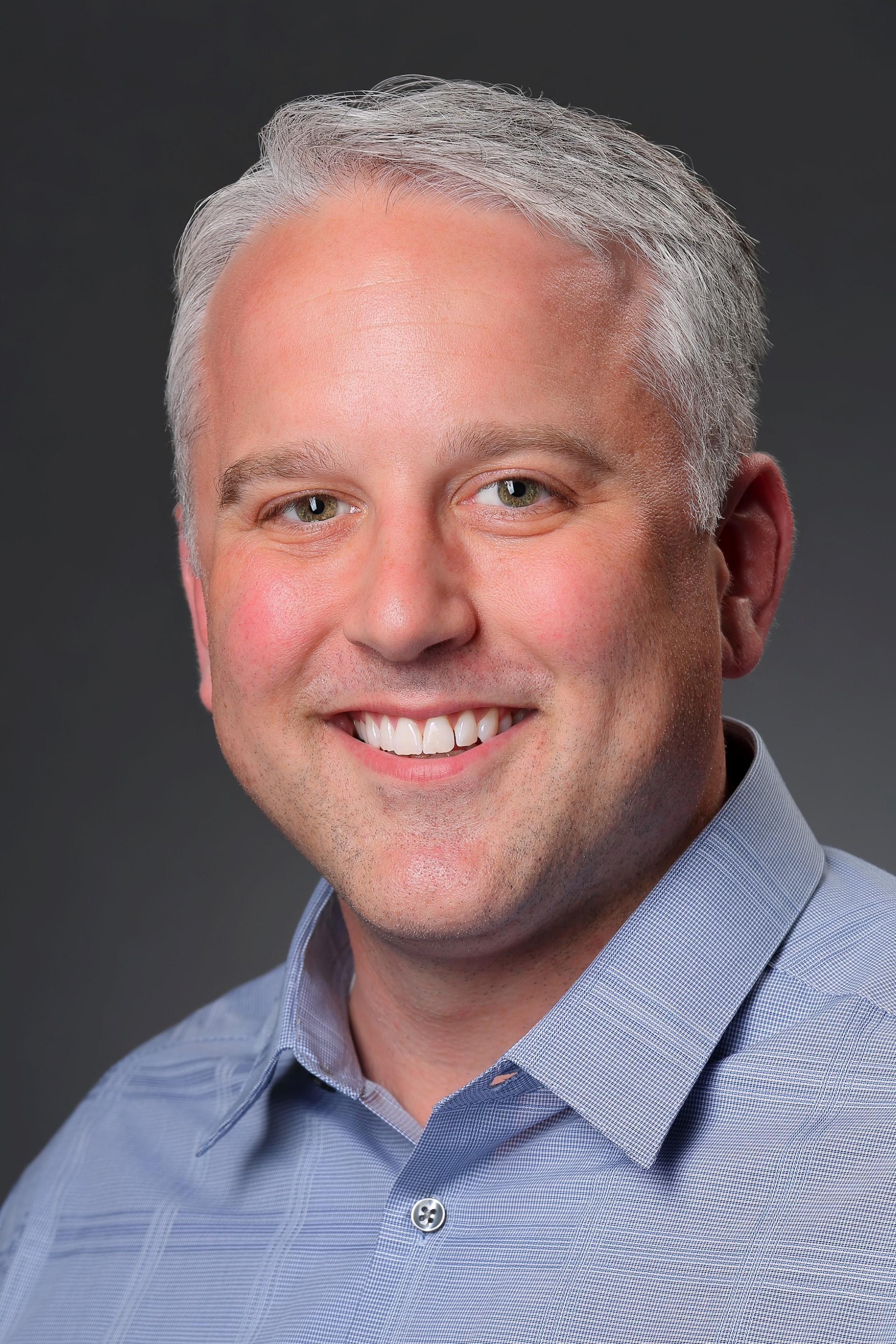
Kevin S. Stepaniuk, DVM, DAVDC, Co-Owner, Pet Dental Specialists, Vancouver, WA
Dr. Stepaniuk has an undergraduate degree from the University of Calgary, DVM degree from Oklahoma State University and completed a small animal internship at Washington State University. His specialty training was in private practice. He is a previous faculty member and section chief of Dentistry and Oral Surgery at the University of Minnesota. He is an invited international and national renowned lecturer and instructor. Dr. Stepaniuk is a journal and textbook author as well as a reviewer for various dentistry and oral surgery publications. He is past-Executive Board member of the AVDC, and past-president of the AVDS. Dr. Stepaniuk works clinically full-time and provides continuing education to veterinarians, veterinary students, and technicians.
Seminar Day-Thursday, September 19th
Practical Veterinary Dentistry to Immediately Implement in Practice
This seminar will be presented with predominately clinical photographs and radiographs. Step-by-step surgical extractions of the maxillary molars in the dog, oronasal fistula repair, and surgical extractions in the cat will be covered. A photographic and radiographic tour of subtle, but commonly missed, clinical examination findings will be presented. What is and what is not feline “stomatitis” to direct the correct diagnosis and treatment plan will be reviewed with updated published literature. Clinical photographs will be used to help participants understand what gingivostomatitis and is not! Fractured teeth and bonded sealants will be presented to correctly understand when and when not they can be considered for use in practice.
8:00 am-8:30 am
Onsite attendees: Registration and Continental Breakfast
Online attendees: Log into WebEx
8:30 am-9:30 am- Oronasal Fistula Repair – Don’t Make It Difficult!
A step-by-step presentation with clinical photographs will be presented for identifying and treating common maxillary canine tooth oronasal fistulas in the dog. A single buccal mucoperiosteal technique will be presented to teach the participant the key principles to have a successful repair with the first surgery. Recognition, pathophysiology, treatment principles, and post-operative care will be outlined for success. At the end of this session participants will be able to: 1. Recognize inapparent and apparent oronasal fistulas of the maxillary canine teeth. 2. Explain how to be successful with the single buccal mucoperiosteal and keep surgical techniques simple for a repair. 3. List the key recommendations such as width of the flap for the defect, tension releasing mucoperiosteal flap, suture technique, and post-operative home care. 4. Discuss how the first attempt at repair is the best attempt at repair and more advanced techniques are rarely needed.
9:30 am-10:30am-Fractured Teeth/Bonded Sealant Recommendations in General Practice
An intraoral photographic and radiographic presentation of fractured teeth, dentin-pulp complex, and treatment recommendations for the AVDC tooth fracture classification will be present for the general practice. Understanding the types of tooth fractures and treatment recommendations and recommended treatments will be reviewed. Bonded sealants in general practice to increase the level of dentistry care that can be provided to the primary care patients will be introduced. At the end of this session participants will be able to: 1. Explain the tooth dentin-pulp complex. 2. Identify the various classifications of tooth fractures and severity. 3. Describe treatment options available for the various tooth fractures beyond extraction and wishful “watching” of a tooth. 4. Describe the roentgen signs of endodontic disease. 5. Discuss information of introducing bonded sealants to their primary care practice.
10:30 am-10:45 am-Break
10:45 am-12:15 pm-Step-By-Step Feline Surgical Extractions
A step-by-step presentation with clinical photographs will be presented for surgical extraction of the feline dentition. The steps for successful extraction will be presented. Pitfalls and pearls in the process will be discussed. The participant will understand how proper equipment can make the process less difficult, more rewarding, and more effective. The regional anatomy will be reviewed to prevent iatrogenic trauma. Participants will learn techniques to close the extraction sites. The common complications reported with feline exodontics will be presented. The prevention of these complications with proper technique will be presented. Management of these complications will be reviewed to help the participant understand how to manage them during the surgical event. At the end of this session participants will be able to: Describe surgical extraction techniques and surgical instrumentation to make extractions less difficult. 2. Identify and respect the vital regional anatomy to avoid iatrogenic trauma. 3. Explain techniques to surgically close the surgical extraction sites. 4. Discuss retained tooth roots and retrieval of retained tooth roots. 5. Distinguish iatrogenic fractures. 6. Treat maxillary lip entrapment and management. 7. Give examples of intraoperative hemorrhage, iatrogenic trauma, pyogenic granuloma, and avoidance.
12:15 pm-1:00 pm-Lunch
1:00 pm-2:00 pm-Clandestine Common Dental Pathology Missed
An intraoral photographic and radiographic presentation of subtle clinical signs of common hidden dental disease that may be easily missed in general practice will be presented. Identification of these subtle signs and lesions in the conscious oral examination should lead the practitioner to recommend intraoral radiographs and treatment for the patient to eliminate chronic infection, inflammation, and pain. Participants will also learn to identify subtle clinical findings during the anesthetized examination. The significance of the pathology will be discussed, and recommended treatments presented. Immediate take home knowledge is learned. At the end of this session participants will be able to: 1. Describe subtle, but common, hidden oral pathology easily missed in the conscious oral examination. 2. Identify hidden dental pathology that can be missed without a meticulous anesthetized examination and intraoral radiographs. 3. List treatment recommendations that should be made after identifying these lesions. 4. Explain the complications to the patient when these diseases go unnoticed and treated
2:00 pm-3:00 pm-Surgical Extraction and Closure of Dog Maxillary Molars Step-By-Step
A step-by-step presentation with clinical photographs will be presented for surgical extraction of the maxillary 4th premolar and maxillary 1st and 2nd molars in the dog. The steps for successful extraction will be presented. Pitfalls and pearls in the process will be discussed. The participant will understand how proper equipment can make the process less difficult, more rewarding, and more effective. The regional anatomy will be reviewed to prevent iatrogenic trauma. Participants will learn techniques to close the extraction sites. At the end of this session participants will be able to: 1. Describe surgical extraction techniques and surgical instrumentation to make extractions less difficult. 2. Discuss techniques for removal of the maxillary molars and 4th premolar. 3. Explain wheel and axel technique for the palatal roots of the maxillary molars. 4. Identify and respect the vital regional anatomy to avoid iatrogenic trauma. 5. Describe techniques to surgically close the maxillary molar extraction sites.
3:00 pm-3:15 pm-Break
3:15 pm-4:15 pm-Feline Stomatitis – Do Not Misdiagnose!
The pathophysiology of the plaque biofilm and periodontal disease will be presented photographs, schematics, and intraoral radiographs. Feline oral inflammation and patterns will be reviewed to help the practitioner to better understand the various etiologies and different treatments necessary for the different patterns of “gingivostomatitis”. An intraoral photographic presentation of diagnosis and treatment to correctly treat different inflammatory diseases in the feline oral cavity. Differentiating aggressive periodontitis from true stomatitis (i.e., caudal mucositis) is discussed. What “stomatitis” is, and is not, will be presented so the practitioner can differentiate and choose the correct treatment path. Discussion of, but not limited to, periodontitis, aggressive periodontitis, feline pyogenic granuloma, oral inflammation, and caudal mucositis (immune dysregulated mucositis/stomatitis) will be presented. At the end of this session participants will be able to: 1. Review important periodontal dental anatomy. 2. Describe the pathophysiology of the plaque biofilm that leads to subgingival periodontal disease and the inflammatory response that results in loss of tooth attachment. 3. Explain prevention and treatment of periodontal disease. 4. Identify patterns of feline oral inflammation. 5. Explain how to approach a “stomatitis” feline patient and understand the importance of caudal mucositis with client communication, treatment planning, prognosis, and treatment.
The Academy Seminar is Generously Sponsored by
ADDITIONAL SEMINAR INFORMATION
- Participants will earn 7.0 hours of continuing education.
- Availability to participate onsite (limited) and online
- Breaks and lunch are provided to onsite attendees
- Handouts are provided to all attendees
- Registration changes (onsite to online) must be received 72 hours prior to the seminar. Changes incur a $40 administrative fee.
- Registration cancellations must be received 72 hours prior to the seminar. Cancellation refund less a 10% administrative fee.
-
Disclaimer: All logos, photos, etc. used in this presentation are the property of their respective copyright owners and are used here for educational purposes only. Please do not take photos of any of the slides.
Online Registration Option
Attend the Seminar, which will begin at 8:00 am (central), from anywhere you have an internet connection! Choose the "online" option during registration and you will be emailed a secure link to attend the live session on the day of the seminar. Only the registrant has access with this link as the attendees are monitored and must sign-in with their name and email address. The registrant is also the only one eligible for the CE credits associated with the Seminar.
What will you experience: Once you have accessed the Seminar your computer screen will be dominated with a direct-feed PowerPoint for clarity of viewing, plus the audio of the presenter as the slides automatically advance on your screen. A small video window on the side of your screen will show the presenter. During the live Q&A session you are able to submit questions.
SEMINAR LOCATION
Minnesota Humanities Center
987 Ivy Avenue East
St. Paul, MN 55106
Seminar Costs
Early-bird registration fee
(now thru August 8th, 2024) |
|
Regular Registration fees
(August 8th-September 5th, 2024) |
|
| MVMA Member / Non-MN DVM |
$195 |
MVMA Member / Non-MN DVM |
$220 |
| MVMA Life / Non-Practicing DVM / Recent Graduate / Graduate Student Intern or Resident |
$130 |
MVMA Life / Non-Practicing DVM / Recent Graduate / Graduate Student Intern or Resident |
$155 |
| Technician / Staff / Other |
$125 |
Technician / Staff / Other |
$150 |
| Non-Member MN Licensed DVM |
$245 |
Non-Member MN Licensed DVM |
$270 |
| DVM Student |
$80 |
DVM Student |
$95 |
If space is still available after September 5th, 2024 prices will increase by $50.
|